Exploring Laminitis
- photos that help unravel the mystery
 |
NEW in November 2023: Revised Exploring Laminitis, with an additional four pages and extra colourization for further insight. Photos of the internal tissues helps us to understand the impact this condition has on both the internal and external aspects of the hoof. These photos open our eyes and minds to the devastation that can occur and allow us to determine when and where we, as professional hoof care providers can help rebuild the compromised hoof. Taking care of the laminitic horse is tough! |
Laminitic hooves do not grow "normally" and the photographic evidence shows us why. This helps us make informed decisions which benefits not only our clients and their horses but also our businesses.
Understanding the underlying anatomy helps us figure out what is actually happening and provides inspiration for ways to help these compromised horses.
Laminitis is rarely just a hoof issue, but as professional hoof care providers, the hoof is our primary concern. We witness the sometimes radical changes to the hoof capsule and know that these changes are not good. But why does the hoof distort so badly? How does this happen?
As farriers, we want to understand the mechanics of laminitis. We need to make sense of the dynamic between the internal and external hoof and how these two components affect each other. Current imaging methods (digital radiographs, MRIs, etc.) answer some of our questions, but to actually see the soft tissues helps us get our heads around how they work together.
We couldn't find this information anywhere and so started dissecting and as a result discovered and photographed a goldmine of information lying just beneath the surface of the hoof. We put this information together into a world class package, Exploring Laminitis.
Exploring Laminitis uses comparative anatomy in a quest to analyze the differences between a "healthy" hoof and one with a hoof with severe, chronic laminitis with capsular rotation. This is part one of a future series that will highlight the different types of laminitic hooves and show the variations that can occur.
We cover some of it here, but you are going to want your own copy to study! The pictures are amazing and especially helpful for visual/kinesthetic learners. There is so much information that it will blow your mind!
The Book takes you on a tour through the layers of the hooves starting with the outside markers
- Compare the external hoof photo with lateral radiographs
- Unique dissections highlight the distortion of the laminitic hoof
- Analyze the displacement of the corinary corium between the hooves
- Investigate the disorganized laminae
- Study the difference in the location of the live sole
- Understand how a bruise caused by the coffin migrates to the surface
- Examine the stretched white line from the inside
- Observe how a ski tip begins to fracture off P3
- Contemplate how bone loss affects the ability of the hoof to regenerate
- Plus much, much more when you engage your mind and explore for yourself!
 |
The new first page compares venograms of two hooves, one with healthy blood flow and one with compromised blood flow. The term 'healthy' is used very loosely in this book. We specifically chose a hoof that looked suspect, but did not have internal changes. | |
 |
In this new spread we show the circumflex artery and two hooves that have ischemia (restricted blood flow). One has regional ischemia and one has perfuse ischemia. | |
 |
Another new page highlights the different types of rotation using both radiographs and diagrams. This leads on to a page that compares the external views of a healthy foot verses a laminititc foot and highlights the common external distortions that occur when laminitis is present. | |
 |
Using radiographs we show the location and orientation of the coffin bone within each of the hoof capsules on both the healthy hoof and rotated hoof. | |
 |
Next a window is cut out to reveal the laminae and coronary coria. This really highlights the damage and distortion to both structures. We also show a sagittal cut of both of the hooves which illustrates how much distortion occurs to the surrounding structures such as the digital cushion. | |
 |
In this spread we take a closer look at the sagittal cuts and study how bruising migrates from the corium outwards through the sole. | |
 |
Comparing the inside of the hoof wall also shows the significant damage that occurs to the epidermal laminae. | |
 |
Here we can see how the sole corium has migrated up over the edge of the coffin bone which is how the sole also became displaced. | |
 |
The damaged and disorganized laminae also have damage to their ends, the terminal laminae. | |
 |
Finally we compare coffin bones from a reasonably healthy vs a known chronic laminitic foot. The laminitic hoof shows bone loss and remodelling and is on the verge of fracturing at the tip. In addition, we have added images from two other hooves: one that had regional ischemia and one with perfuse ischemia. |
|
|
Printed Book - $40Printed on heavy duty card and spiral bound to make this guide very rugged and easy to use as a portable teaching tool. 6"x11", 18 pages. eBook - $30
|
Testimonials
Hi Paige and Jenny – congratulations on an excellent publication. I thoroughly enjoyed turning the pages and having the labels appear on the following page – a great initiative. The graphics and compositing are first class. I am preparing a new edition of my own book to be retitled The Illustrated Horse Foot. I hope it matches yours in graphical impact. It will be in print first and later as an eBook.
Professor Chris Pollitt BVSc PhD
Got the Laminitis 1 today and WOW!!!!!!! I am beyond impressed. I will now proceed to make half of my clientele cringe, and hopefully make them think a little bit.
Åsa Jahnke Stephens
Subscribe to Anatomy of the Equine News
Stay informed by subscribing to our newsletter and receive a FREE copy of our Distal Limb, Laminae & Digital Cushion eBook as a thank you!


Look for New! bundles
Bundles
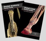 Head to Hoof Printed Pack Buy the Dental Anatomy Guide along with the Distal Limb Guide and save $10 on the combined price and we'll give you a free copy of the Digital Pulse eBook too! Pack price: $40 |
|
Head to Hoof eBook Pack Same books as above but all in eBook format. Price: $25 |
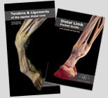 Leg Printed Pack Buy the Tendon & Ligament Guide along with the Distal Limb Guide and save $10 on the combined price and we'll give you a free copy of the Coffin Bone eBook too! Pack price: $50 |
|
Leg eBook Pack Same books as above but all in eBook format. Price: $35 |
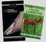 Laminitis Pack Buy Exploring Laminitis along with the Equine Laminitis Guide and save $10 on the combined price and we'll give you a free copy of the Digital Pulse eBook too! Pack price: $40 |
|
Laminitis eBook Pack Same books as above but all in eBook format. Price: $28 |
NEW!
Everything Pack
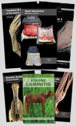
Buy all six books including the new Hoof Distortions book: Hoof Distortions; Tendon & Ligament Guide; Distal Limb Guide; Exploring Laminitis; Equine Laminitis Guide; and Dental Anatomy Guide and save $30 on the combined price and we'll give you a free copy of the Coffin Bone eBook ($10 value) and Digital Pulse eBook ($3 value) too!
Pack price: $150
NEW!
Everything eBook Pack
Same books as above but all in eBook format.
Price: $100
Poster
Bundles
|
Back in Stock!  Buy a set of SEVEN posters and save $10 per poster. Pack price: $70 |
What is an eBook?
An eBook is an electronic book that is compatible with any computer, laptop, smart phone or tablet. When you order the eBook you will be sent an email with a link to the download file - click on that link and make sure you save it to a place where you can find it again!